Overview Achilles Tendon Rupture Surgery is a surgical procedure is used to repair a ruptured Achilles tendon. There are two types of surgery an open surgery and a percutaneous surgery, in both cases the surgeon sews the tendon back together through the incision helping the tendon to heal properly and restore function to the foot and ankle. If the injury or rupture has caused severe swelling the surgery may be delayed for a short period of time to allow the swelling to go down. Causes Achilles tendon ruptures are most likely to occur in sports requiring sudden stretching, such as sprinting and racquet sports. Achilles tendon ruptures can happen to anyone, but are most likely to occur to middle age athletes who have not been training or who have been doing relatively little training. Common sporting activities related to Achilles tendon rupture include, badminton, tennis, squash. Less common sporting activities that can lead to Achilles tendon rupture include: TKD, soccer etc. Occasionally the sufferer may have a history of having had pain in the Achilles tendon in the past and was treated with steroid injection to around the tendon by a doctor. This can lead to weakening of the tendon predisposing it to complete rupture. Certain antibiotics taken by mouth or by intravenous route can weaken the Achilles tendon predisposing it to rupture. An example would be the quinolone group of antibiotics. An common example is Ciprofloxacin (or Ciprobay). Symptoms Whereas calf strains and tendonitis may cause tightness or pain in the leg, Achilles tendon ruptures are typically accompanied by a popping sensation and noise at the time of the injury. In fact, some patients joke that the popping sound was loud enough to make them think they?d been shot. Seeing a board-certified orthopedic surgeon is the best way to determine whether you have suffered an Achilles tendon tear. Diagnosis A diagnosis can be made clinically, but an MRI or ultrasound scan can confirm it. On examination, the patient will present with reduced plantarflexion strength, a positive Thompson test and potentially, a palpable gap in the Achilles. The whole length of the tendon should be examined to check for injuries that can occur at the insertion and the musculotendinous junction. Non Surgical Treatment This condition should be diagnosed and treated as soon as possible, because prompt treatment probably improves recovery. You may need to be referred urgently to see a doctor in an orthopaedic department or accident and emergency department. Meanwhile, if a ruptured Achilles tendon is suspected, you should not put any weight on that foot, so do not walk on it at all.Treatment options for an Achilles tendon rupture include surgical and non-surgical approaches. The decision of whether to proceed with surgery or non-surgical treatment is based on the severity of the rupture and the patient?s health status and activity level. Non-surgical treatment, which is generally associated with a higher rate of re-rupture, is selected for minor ruptures, less active patients, and those with medical conditions that prevent them from undergoing surgery. Non-surgical treatment involves use of a cast, walking boot, or brace to restrict motion and allow the torn tendon to heal. 
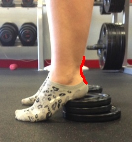
Surgical Treatment Some surgeons feel an early surgical repair of the tendon is beneficial. The surgical option was long thought to offer a significantly smaller risk of re-rupture compared to traditional non-operative management (5% vs 15%). Of course, surgery imposes higher relative risks of perioperative mortality and morbidity e.g. infection including MRSA, bleeding, deep vein thrombosis, lingering anesthesia effects, etc.
Overview
The condition of flat feet in adults is known as ?fallen arches.? Not all adults develop flat feet, and some people are more prone to developing the condition than others. An obese person puts extra weight on their feet while walking or standing. Over time, this can weaken the components that make up the arch and cause the arch to collapse. A woman who is pregnant may also suffer from flat feet during her pregnancy. The problem with developing flat feet as an adult is that in most cases the changes are permanent, if not bothersome. Doctors recommend using custom-made orthotics in shoes to treat the problem. Flat feet were once considered a result of poor health, but it has been proven that athletes such as runners, who are in great condition, also suffer from flat feet. In fact, it?s very common among track runners. Flat feet were once thought of as a bad thing. But studies show that people with higher arches are four times more likely to injure or sprain their ankles than people with flat feet. Studies conducted by the military have discredited the idea that flat feet are a reason to be excused from service. Causes As discussed above, many health conditions can create a painful flatfoot. Damage to the posterior tibial tendon is the most common cause of AAFD. The posterior tibial tendon is one of the most important tendons of the leg. It starts at a muscle in the calf, travels down the inside of the lower leg and attaches to the bones on the inside of the foot. The main function of this tendon is to hold up the arch and support your foot when you walk. If the tendon becomes inflamed or torn, the arch will slowly collapse. Women and people over 40 are more likely to develop problems with the posterior tibial tendon. Other risk factors include obesity, diabetes, and hypertension. Having flat feet since childhood increases the risk of developing a tear in the posterior tibial tendon. In addition, people who are involved in high impact sports, such as basketball, tennis, or soccer, may have tears of the tendon from repetitive use. Symptoms Feet tire easily and become painful and achy, especially around the arch, ankle and heel. Swelling on the inside bottom of your feet. Back and leg pain. Difficulty standing on toes. Diagnosis It is important for people with foot pain to know if they have flat feet. The following tests can help you determine your arch type. When you get out of a swimming pool, look at your footprint on the concrete. The front of the foot will be joined to the heel by a strip. If your foot is flat, then the strip is the same width as the front of the foot, creating a footprint that looks like a stretched out pancake. With a normal arch, the strip is about half the width of the front of the foot. If you have a high arch, only a thin strip connects the front of the foot with the heel. Put your shoes on a flat table and view them at eye level from behind. See if the sole is worn evenly. A flat foot will cause more wear on the inside of the sole, especially in the heel area. The shoe will easily rock side to side. A flat foot will also cause the upper part of the shoe to lean inward over the sole. Both shoes should wear about the same way. If you have pain in one foot, you should make sure you don't have a fallen arch on that side. There are two good tests you can perform at home to detect this problem. Place your fingertips on a wall that you are directly facing and stand on your tiptoes on one foot. If you can't do it, a fallen arch may be the culprit. Stand with your feet parallel. Have someone stand in back of you and look at your feet from behind. You can also do it yourself if you stand with your back to a mirror. Normally, only the pinky toe is visible from behind. If one foot is flatter than the other, the 4th and sometimes the 3rd toe on that foot can also be seen. Non Surgical Treatment If your condition is bothersome, try elevating your feet and using ice on the arches to reduce swelling. Your podiatrist can recommend several orthotic aids and inserts to strengthen the tendons of your foot. He can also demonstrate stretching exercises or refer you to physical therapy to get those tendons back into shape. If the symptoms of fallen arches are painful and troubling, he may recommend a steroid injection to relieve inflammation and pain. And in some instances, he may determine that surgery is necessary. Surgical Treatment A combination of surgical procedures can be used to reconstruct the flatfoot. Generally, these procedures can be separated into those that correct deformities of the bones and those that repair ligaments and tendons. Your orthopaedic surgeon will choose the proper combination of procedures for your foot. Surgery of the foot can be performed under regional anesthesia, which is numbing the foot and ankle with a nerve or spinal block, or general anesthesia, which may require a breathing tube. A nerve block is often placed behind the knee to reduce pain after surgery. Overview
Each foot contains 26 bones, which form two arches. The longitudinal arch runs the length of the foot, and the transverse arch runs the width. The bones of the arch are primarily held together by the shape with which they fit with each other and by fibrous tissues known as ligaments that serve to hold the bones to each other. The muscles of the foot, along with a tough, sinewy tissue known as the plantar fascia, provide secondary support to the foot. There are also fat pads in the foot to help with weight-bearing and absorbing impact. Arch pain can occur whenever something goes wrong with the function or interaction of any of these structures. Causes The number one cause of arch pain is Plantar Fasciitis, and you'll be glad to know that more than 90% of cases of this painful condition can be resolved with simple, conservative at-home treatments. While extremely severe cases of Plantar Fasciitis may require cortisone injections or surgeries, most people can experience quick relief and eventual recovery with the right combination of non-invasive therapies. Symptoms The foot of a newborn with congenital vertical talus typically has a convex rocker-bottom shape. This is sometimes combined with an actual fold in the middle of the foot. The rare person who is diagnosed at an older age often has a "peg-leg" gait, poor balance and heavy calluses on the soles where the arch would normally be. If a child with congenital vertical talus has a genetic disorder, additional symptoms often are seen in other parts of the body. Diagnosis The diagnosis of high arch (cavus) foot deformity or Charcot Marie Tooth disease can be made by an orthopedic surgeon in the office. Evaluation includes a thorough history and physical examination as well as imaging studies such as X-rays. The orthopedic surgeon will look at the overall shape, flexibility, and strength of a patient?s foot and ankle to help determine the best treatment. Nerve tests may occasionally need to be performed to help confirm the diagnosis. Non Surgical Treatment Anti-inflammatory medications such as ibuprofen and aspirin can provide short-term relief from foot pain associated with fallen arches, notes the American Academy of Orthopaedic Surgeons. Doctors sometimes inject the foot with a corticosteroid medication, which acts as an anti-inflammatory, to relieve acute pain. Although medications can provide symptom relief, they do not correct the underlying foot abnormality. Surgical Treatment Cavus foot is caused in part by an over-pull of one of the lateral ankle muscles. A release of this tendon can be performed on the outside of the ankle. Additionally, a transfer of this tendon can be performed to help in correcting deformity of the ankle joint. Often patients will have a tightness of their gastrocnemius muscle, one of the main muscles in the calf. This can increase the deformity or prevent a correction from working. It is addressed with a lengthening of a part of the calf muscle or Achilles tendon. This is often performed through one or more small cuts in the back of the leg or ankle. Finally, the plantar fascia may be tight. The plantar fascia is a cord-like structure that runs from the heel to the front part of the foot. Partial or complete plantar fascia release may be done. Prevention Foot and ankle injuries are common in sports, especially running, tennis and soccer. But sports enthusiasts can decrease the risk of injury by taking some precautions. Lightly stretch or better yet, do a slow jog for two to three minutes to warm up the muscles. Don't force the stretch with a "bouncing motion." The amount of time spent on the activity should be increased gradually over a period of weeks to build both muscle strength and mobility. Cross training by participating in different activities can help build the muscles. People whose feet pronate or who have low arches should choose shoes that provide support in both the front of the shoe and under the arch. The heel and heel counter (back of the shoe) should be very stable. Those with a stiffer foot or high arches should choose shoes with more cushion and a softer platform. Use sport-specific shoes. Cross training shoes are an overall good choice; however, it is best to use shoes designed for the sport. Stretching Exercises Calf Raises. Strengthens the tendons in your heels and calf muscles, which support your arch. Raise up on the balls of your feet as high as possible. Slowly lower down. Do three sets of 10 reps. Progress to doing the raises on stairs (with heels hanging off), and then to single-leg raises. Step Stretch. Improves flexibility in your Achilles tendon and calf-when these areas become tight, the arch gets painfully overloaded. Stand at the edge of a step, toes on step, heels hanging off. Lower your heels down, past the step, then raise back up to the start position. Do three sets of 10 reps. Doming. Works the arch muscles and the tibialis posterior (in the calf and foot) to control excess pronation. While standing, press your toes downward into the ground while keeping the heel planted, so that your foot forms an arch (or dome). Release, and do three sets of 10 reps on each foot. Toe Spread and Squeeze. Targets the interossei muscles of the foot, which support the arch. While sitting, loop a small resistance band around your toes. Spread toes; release. Then place a toe separator (used at nail salons) in between toes. Squeeze toes in; release. Do three sets of 10 reps of each exercise on both feet. Towel Curls. Works the toe-flexor muscles that run along your arch to increase overall foot strength. Lay a small hand towel on the floor, and place one foot on the towel. Using just your toes, scrunch the towel toward you, hold, then slowly push the towel away from you back to start position. Do three sets of 10 reps on each foot. Overview
Pes planus is the scientific term that describes low arch or "flat" feet. As podiatric physicians, we are very interested not only in structure but also in function of the feet. While very few people have 20-20 vision, it is also true that very few people have perfect arch structure. High and low arch feet are just the two ends of the spectrum of foot structure. The more deviation from what is considered perfect, the worse the function becomes. We describe deviation from "the ideal" as imbalance in structure. It is the imbalance in structure that leads to abnormal function. Abnormal function causes pain and/or deformity. Deformity can manifest itself in a variety of foot problems such as bunions or hammertoes. Likewise, pain can manifest itself in a variety of ways such as heel pain ( plantar fasciitis or heel spur), corns or calluses, metatarsalgia or pain in the ball of the foot ( neuromas, stress fractures, or tendonitis), or even in pediatric problems. Invariably, we can treat most foot imbalance or biomechanical insufficiencies with orthotic therapy. Causes Arch pain is most often caused by plantar fasciitis, which can affect the heel, arch, or both. Plantar fasciitis treatment is the same, regardless of the location of foot pain (see above). For persistent plantar fasciitis, an injection with a mixture of a steroid and local anesthetic can be helpful. Fallen arches or flat feet occur when the arches of the feet flatten out (often when standing or walking), causing foot pain and other problems. Flat feet can be treated with shoe inserts (orthotics), shoe adjustments, rest/ice, using a walking cane or brace, or physical therapy. Occasionally, surgery is necessary. Symptoms The muscle imbalance around the foot and ankle gives rise to a typical pattern of deformity in addition to the high arch (known as cavus). The bone under the big toe (called the first metatarsal) can become very prominent and the toes can curl or clench like a fist (called claw toes). Excessive amount of weight may be placed on the ball and heel of the foot, which can lead to the ankle weakening and giving way (this is referred to as ankle instability) and soreness. Calluses and sometimes stress fractures may occur where the foot is exposed to extra friction or pressure, such as on the outer (or lateral) border of the foot. Diagnosis In people with flat feet, the instep of the foot comes in contact with the ground when standing. To diagnose the problem, the health care provider will ask you to stand on your toes. If an arch forms,the flat foot is called flexible. You will not need any more tests or treatment. If the arch does not form with toe-standing (called rigid flat feet), or if there is pain, other tests may be needed, including a CT scan to look at the bones in the foot. MRI scan to look at the tendons in the foot. X-ray of the foot. Non Surgical Treatment If you have arch pain, you need proper arch support. You can get arch support by purchasing custom shoe inserts that are made to support your feet. If you have flat feet or high arches, you can certainly benefit from arch support inserts. Take a look at your wet footprint; if you notice that your footprint is completely filled in, then you have flat feet. On the other hand, if there is a large crescent shape missing from your footprint, then you have high arches. Both of these conditions require proper support from a shoe insert. Foot Solutions You can also take care of your feet by avoiding high heels and flip-flops. If you must wear high heels, choose a heel that is two inches or less, and try to wear them only for short periods of time. Flip-flops provide very little support, so wear them only if you won?t be doing very much walking. Surgical Treatment In adults, the most common cause of collapse is due to the posterior tibial tendon tear. In such cases, the tendon must be repaired and a second tendon may be added to the posterior tibial tendon for strength and added support. If the foot is found to be very flat, bone realignment procedures or possible bone fusion procedures may be used to realign the foot. If the calf or Achilles tendon are found to be tight, they may be lengthened to allow better motion at the ankle and less arch strain. The forefoot may also be in a poor position and stabilization of the arch may be necessary to increase forefoot contact to the ground. Prevention People who run regularly should replace shoes every six months, more frequently if an avid runner. Avoid running or stepping on uneven surfaces. Try to be careful on rocky terrain or hills with loose gravel. Holes, tree stumps and roots are problems if you are trail running. If you have problems with the lower legs, a dirt road is softer than asphalt, which is softer than concrete. Try to pick a good surface if possible. However, if you're racing, be sure to train on the surface you'll eventually run on. Be careful running too many hills. Running uphill is a great workout, but make sure you gradually build this up to avoid injuries. Be careful when running downhill too fast, which can often lead to more injuries than running uphills. Prevent recurrent injuries. Athletes who have experienced ankle injuries previously may benefit from using a brace or tape to prevent recurrent ankle injuries. Stretching Exercises Achilles stretch. Stand with the ball of one foot on a stair. Reach for the step below with your heel until you feel a stretch in the arch of your foot. Hold this position for 15 to 30 seconds and then relax. Repeat 3 times. Balance and reach exercises. Stand next to a chair with your injured leg farther from the chair. The chair will provide support if you need it. Stand on the foot of your injured leg and bend your knee slightly. Try to raise the arch of this foot while keeping your big toe on the floor. Keep your foot in this position. With the hand that is farther away from the chair, reach forward in front of you by bending at the waist. Avoid bending your knee any more as you do this. Repeat this 15 times. To make the exercise more challenging, reach farther in front of you. Do 2 sets of 15. While keeping your arch raised, reach the hand that is farther away from the chair across your body toward the chair. The farther you reach, the more challenging the exercise. Do 2 sets of 15. Towel pickup. With your heel on the ground, pick up a towel with your toes. Release. Repeat 10 to 20 times. When this gets easy, add more resistance by placing a book or small weight on the towel. Resisted ankle plantar flexion. Sit with your injured leg stretched out in front of you. Loop the tubing around the ball of your foot. Hold the ends of the tubing with both hands. Gently press the ball of your foot down and point your toes, stretching the tubing. Return to the starting position. Do 2 sets of 15. Resisted ankle dorsiflexion. Tie a knot in one end of the elastic tubing and shut the knot in a door. Tie a loop in the other end of the tubing and put the foot on your injured side through the loop so that the tubing goes around the top of the foot. Sit facing the door with your injured leg straight out in front of you. Move away from the door until there is tension in the tubing. Keeping your leg straight, pull the top of your foot toward your body, stretching the tubing. Slowly return to the starting position. Do 2 sets of 15. Heel raise. Stand behind a chair or counter with both feet flat on the floor. Using the chair or counter as a support, rise up onto your toes and hold for 5 seconds. Then slowly lower yourself down without holding onto the support. (It's OK to keep holding onto the support if you need to.) When this exercise becomes less painful, try doing this exercise while you are standing on the injured leg only. Repeat 15 times. Do 2 sets of 15. Rest 30 seconds between sets. Overview
Adult acquired can end up being a extremely severe situation that can lead to many secondary deformities, not only inside the particular foot but additionally in the knees, hips and also back. This kind of presentation discusses a brand name new scientifically verified procedure that might be able to assist realign and connect this challenge in its root. Causes Several risk elements are generally associated with PTT dysfunction, which includes high blood pressure, obesity, diabetes, previous ankle surgery or perhaps trauma as well as exposure to become able to steroids. A New one who suspects that they are usually suffering via PTT dysfunction ought to seek health-related focus earlier rather than later. This is much simpler to treat early and steer distinct of a new collapsed arch laptop or computer is often to repair one. Whenever the pain 1st happens there is no significant flatfoot deformity, original remedies contain rest, oral anti-inflammatory medications and, depending around the severity, a unique boot or brace. Symptoms As different forms of flatfoot get distinct causes, the associated signs and symptoms may vary for different people. Several generalized signs and symptoms are usually listed. Pain over the span of your posterior tibial tendon which in turn lies on the inside in the foot and ankle. This specific could be related using swelling on the inside with the ankle. Pain that's more serious using activity. Higher intensity or perhaps impact activities, for example operating and also jumping, can always be extremely difficult. A Few patients can easily have difficulties walking or even standing for long intervals of your own time and may encounter pain from the interior with the ankle along with in the arch in the foot. feeling just like one is ?dragging their own foot.? When the foot collapses, the heel bone may shift position and set stress about the outside ankle bone (fibula). This specific may cause pain in the bones and tendons within the outside of the ankle joint. Patients together with a vintage injury or even arthritis inside the middle involving the foot may have got painful, bony bumps around the top along with inside of the foot. These types of create shoe put on really difficult. Sometimes, the bony spurs are usually consequently big they pinch the particular nerves that can lead to numbness as well as tingling about the top of the actual foot and also in to always be able to the toes. Diabetic patients may well not expertise pain if they've got damage to their nerves. That They may only notice swelling as well as a large bump about the bottom with the foot. The Particular big bump could cause skin problems as well as an ulcer (a sore that doesn't heal) could develop if appropriate diabetic shoe don isn't used. Diagnosis Observation with a skilled foot clinician and a hands-on evaluation with the foot as well as ankle may be the nearly all accurate diagnostic technique. Your Own Dallas foot doctor may get you do a new strolling examination (the most dependable way to examine for that deformity). during walking, the affected foot appears much more pronated along with deformed. Your Current podiatrist may do muscle testing to appear with regard to power deficiencies. Throughout the single foot raise test, the actual foot physician will have you rise up around the suggestion of one's toes although trying for you to keep the unaffected foot off the particular ground. In case your posterior tendon has been attenuated or perhaps ruptured, anyone will be unable to lift your current heel off the particular floor. Inside less severe cases, it is possible to rise on your current toes, nevertheless the heel will not invert normally. X-rays are usually not necessarily beneficial as a diagnostic tool for Adult Flatfoot simply because each feet will usually demonstrate any deformity. MRI (magnetic resonance imaging) could show tendon injury and inflammation, but can?t always be relied upon for any complete diagnosis. in the majority of cases, a MRI is not necessary to diagnose any posterior tibial tendon injury. An ultrasound might also become used to confirm the particular deformity, however is usually not necessary for an original diagnosis. Non surgical Treatment Initial treatment method for most patients consists associated with rest and also anti-inflammatory medications. This kind of will help reduce the swelling as well as pain connected with the condition. The Actual long lasting remedy for the issue typically involves custom made orthotics and supportive shoe gear in order to avoid further breakdown of the foot. ESWT(extracorporeal shock wave therapy) can become a novel therapy which in turn utilizes sound wave technology to become able to stimulate blood flow for the tendon to accelerate your healing process. This particular might help cause an even more rapid return to regular actions for the majority of patients. In case treatment method will be initiated early within the process, many patients can easily experience the come back again to typical activities with out your requirement for surgery. Surgical Treatment The indications regarding surgery tend to be persistent pain and/or significant deformity. Sometimes the foot just feels weak and the assessment involving deformity is better carried out with a foot and ankle specialist. In case surgery can be appropriate, the combination of soft tissue along with bony processes might always be considered in order to appropriate alignment and also support the medial arch, taking strain off failing ligaments. Based upon your tissues involved and extent of deformity, the particular foot as well as ankle specialist will determine the particular necessary mix of procedures. Surgical methods could include a medial slide calcaneal osteotomy for you to right place with the heel, a new lateral column lengthening to always be able to right position within the midfoot plus a medial cuneiform osteotomy or initial metatarsal-tarsal fusion in order to correct elevation with the medial forefoot. Your posterior tibial tendon may be reconstructed having a tendon transfer. Inside severe cases (stage III), the actual reconstruction may include fusion with the hind foot,, causing stiffness of the hind foot however the desired pain relief. in probably your most severe stage (stage IV), the particular deltoid ligament on the inside in the ankle fails, resulting in the deformity inside the ankle. This particular deformity over time can lead to arthritis inside the ankle. Overview
Just as an old bridge would grow to become able to be unsafe because the construction was compromised, the feet may feel the same. If you have PTTD as well as the tendon will carry on to weaken, it simply won?t have the actual ability maintain your arch. Throughout this situation, the particular arch can easily fall or collapse, that is referred in order to as adult-acquired flat foot. you might encounter pain along with tenderness powering the inside with the ankle. over time, additionally to the flattening in the foot, anyone may also recognize a new misalignment of the heel so that it will no longer aligns underneath the remainder of the leg. The tightening in the heel cord, arthritis, and also achievable deformity with the ankle joint could also occur. An Individual might glance at the discomfort move from the inside towards the not inside the ankle as well. Flat foot could in addition contribute to problems using the plantar fascia, which is the actual tendon that runs along the duration of your arch. Causes Causes of a grown-up acquired flatfoot can include Neuropathic foot (Charcot foot) secondary to end up being able to Diabetes mellitus, Leprosy, Profound peripheral neuritis involving any cause. Degenerative changes in the ankle, talonavicular as well as tarsometatarsal joints, or both, secondary in order to Inflammatory arthropathy, Osteoarthropathy, Fractures, Acquired flatfoot resulting coming from loss in the supporting structures in the medial longitudinal arch. Dysfunction of the tibialis posterior tendon Tear of the spring (calcaneoanvicular) ligament (rare). Tibialis anterior rupture (rare). Painful flatfoot can have various other causes, for example tarsal coalition, however as this kind of affected person is not going to present using a change within the model of your foot these aren't included here. Symptoms Pain along the inside of the foot along with ankle, where the tendon lies. This specific could or even may not necessarily be related using swelling inside the area. Pain that's more serious along with activity. High-intensity or perhaps high-impact activities, such as running, can always be quite difficult. A Amount Of patients can possess trouble walking or perhaps standing for any extended time. Pain around the outside involving the ankle. When the foot collapses, the actual heel bone might shift in order to a new position outwards. This kind of can put stress on the outside ankle bone. The Actual same kind involving pain can be present in arthritis inside the back with the foot. Asymmetrical collapsing of the medial arch about the affected side. Diagnosis Perform a new structural assessment with the foot as well as ankle. Verify the actual ankle with regard to alignment and also position. Whenever it comes down to patients using severe PTTD, the actual deltoid provides failed, causing an instability in the ankle as well as possible valgus of the ankle. This kind of can be a uncommon and challenging problem in order to address. However, if 1 misses it, it could lead to dire consequences along with potential surgical failure. Examine the heel alignment and also place in the heel both loaded along with throughout varus/valgus stress. Compare selection of motion of the heel towards the typical contralateral limb. check alignment of the midtarsal joint pertaining to collapse as well as lateral deviation. Noting your degree of lateral deviation in contrast towards the contralateral limb is important for surgical planning. Verify midfoot alignment in the naviculocuneiform joints along with metatarsocuneiform joints each with regard to sag and hypermobility. Non surgical Treatment Non-surgical treatment consists of rest along with decreasing the exercise until the pain improves. Orthotics or even bracing assist support the tendon to reduce its pull along the arch, thus reducing pain. Inside reasonable to end up being able to severe cases, a new down below knee cast or perhaps strolling boot may be required to enable the tendon to relax entirely and heal. Bodily therapy is an integral section of the non-surgical therapy routine to lessen inflammation along with pain. Anti-inflammatory medication is actually often utilized as well. Several times evaluation regarding your existing shoes can be necessary to ensure you're wearing appropriate shoe gear to avoid re-injury. Surgical Treatment If original conservative therapy of posterior tibial tendon insufficiency fails, surgical treatment method is considered. Operative treatment involving stage one disease involves relieve the tendon sheath, tenosynovectomy, debridement with the tendon together with excision regarding flap tears, along with repair of longitudinal tears. a short-leg strolling cast can be worn pertaining to 3 weeks postoperatively. Teasdall and Johnson reported total relief associated with pain throughout 74% associated with 14 patients undergoing this remedy routine regarding stage 1 disease. Surgical debridement involving tenosynovitis in first stages is considered to be able to probably prevent progression associated with disease for you to later stages regarding dysfunction. Overview 
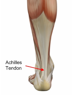
The Achilles tendon is the largest and strongest tendon in the human body. The Achilles tendon connects the heel bone (calcaneus) to the muscles at the back of the calf (using gastrocnemius and soleus muscles). The synchronous function of the tendon and calf muscles is critical for activities like jumping, running, standing on the toe, and climbing stairs. When climbing stairs or running, the forces within the tendon have been measured and indicate that the structure is able to withstand at least 10 times the body weight of the individual. The function of the Achilles tendon is to help raise your heel as you walk. The tendon also assists in pushing up the toes and lifting the rear of the heel. Without an intact Achilles tendon, almost any motion with the ankle (for example, walking or running) is difficult.
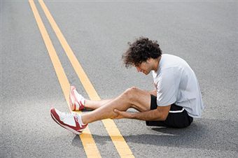
 Causes Achilles tendon rupture occurs in people that engage in strenuous activity, who are usually sedentary and have weakened tendons, or in people who have had previous chronic injury to their Achilles tendons. Previous injury to the tendon can be caused by overuse, improper stretching habits, worn-out or improperly fitting shoes, or poor biomechanics (flat-feet). The risk of tendon rupture is also increased with the use of quinolone antibiotics (e.g. ciprofloxacin, Levaquin). Symptoms Whereas calf strains and tendonitis may cause tightness or pain in the leg, Achilles tendon ruptures are typically accompanied by a popping sensation and noise at the time of the injury. In fact, some patients joke that the popping sound was loud enough to make them think theyd been shot. Seeing a board-certified orthopedic surgeon is the best way to determine whether you have suffered an Achilles tendon tear. Diagnosis The actual area of an Achilles tendon rupture cannot be seen on x-ray. Therefore, although x-rays are often done to rule out bony injuries in individuals with an Achilles tendon rupture these x-rays are usually normal. Diagnostic ultrasound of the tendon can be performed to assess the integrity of the tendon. Other diagnostic tests, such as MRI's, may also be required in difficult cases. Non Surgical Treatment Non-surgical treatment of Achilles tendon rupture is usually reserved for patients who are relatively sedentary or may be at higher risk for complications with surgical intervention (due to other associated medical problems). This involves a period of immobilization, followed by range of motion and strengthening exercises; unfortunately, it is associated with a higher risk of re-rupture of the tendon, and possibly a less optimal functional outcome. Surgical Treatment An Achilles tendon rupture is a complete tear of the fibrous tissue that connects the heel to the calf muscle. This is often caused by a sudden movement that overextends the tendon and usually occurs while running or playing sports such as basketball or racquetball. Achilles tendon rupture can affect anyone, but occurs most often in middle-aged men. Prevention Prevention centers on appropriate daily Achilles stretching and pre-activity warm-up. Maintain a continuous level of activity in your sport or work up gradually to full participation if you have been out of the sport for a period of time. Good overall muscle conditioning helps maintain a healthy tendon. |
AuthorWrite something about yourself. No need to be fancy, just an overview. Archives
July 2017
Categories |